Melanoma (malignant melanoma) is a malignant tumor arising from melanocytes present in the skin, mucosa (mucous membrane lining various structures) and occasionally from melanocytes in the gastrointestinal tract, eye, brain or other organs. Melanocytes contain melanin, a dark pigment that produces skin color. Nevi (moles) are benign melanocytic lesions. There are many other melanocytic lesions (benign and malignant), which are listed in the Table of Contents here.
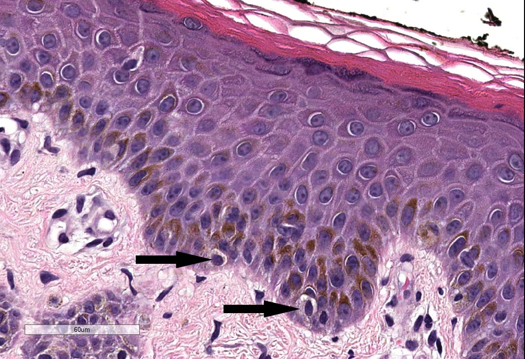
The arrows point to melanocytes in the epidermis, which are typically at the basal (lower) layer. They have a clearing around the nucleus because they lack desmosomes found in other epidermal cells (keratinocytes) which attach these cells to their neighbors and make the skin waterproof and tough. Source: Wikipedia and PathologyOutlines.com.
Melanocytes vs Keratinocytes Made Easy: Source: Jerad Gardner, M.D.
Melanoma causes only 1% of skin cancer cases but most skin cancer deaths. The American Cancer Society estimates that there will be 97,000 new melanoma cases and 8,000 deaths in the U.S. in 2023. Melanoma cases have been increasing in women ages 50+ but have been stable in men. Melanoma deaths have been decreasing due to improved treatment.
Melanoma is usually but not always caused by ultraviolet skin damage. It can be recognized in many cases by the ABCDE rule: Asymmetry, Border irregularity, Color variation, Diameter > 6mm and Evolution.
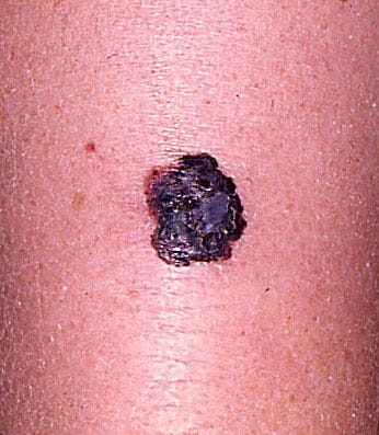
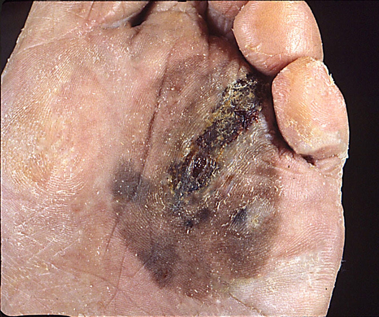
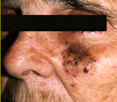
These clinical images of melanoma were contributed by Michele Donati, M.D.
49 year old woman with a 1.5 cm slightly elevated and highly pigmented lesion with irregular borders.
79 year old man with a 6 cm large and irregularly pigmented lesion on the sole of the foot.
82 year old man with a 5.5 cm irregular, pigmented macule with multiple nodular areas on the face.
Melanoma looks different from the other surrounding moles in the same individual (“ugly duckling sign”)
Pigmentation beyond the proximal nail fold is an important clinical clue of subungual (below the nail) melanoma.
Contributed by Julia Nunley, M.D.
Irregular pigmented macule on forearm with chronic actinic damage. Biopsy revealed melanoma in situ (lentigo maligna), which is an early form of melanoma.
Microscopically, melanoma is characterized by tumor cells in the epidermis and dermis.
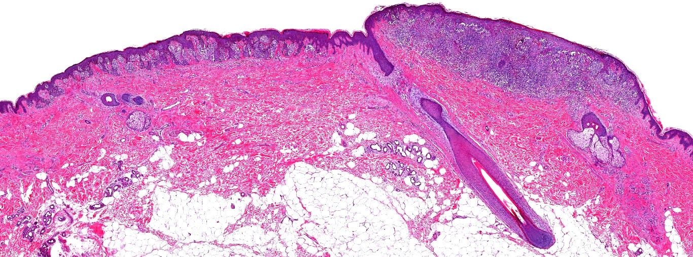
Normal skin has only occasional melanocytes in the epidermis (its layers are labeled). The dermis lacks melanocytes and has few cells. Contributed by Mariantonieta Tirado, M.D.
These microscopic images of melanoma were contributed by Michele Donati, M.D., Jijgee Munkhdelger, M.D., Ph.D. and Andrey Bychkov, M.D., Ph.D.
59 year old woman with a 1.2 cm lesion on the trunk. The lesion (upper right side) is asymmetrical, one of the ABCDEs of melanoma.
44 year old woman with 0.8 cm nodule on her back. The nodule is composed primarily of melanoma cells.
In this melanoma specimen, there are nests of melanoma cells with prominent pigment in the dermis.
56 year old woman with 0.6 cm slightly elevated pigmented lesion on the lower back. This lesion is symmetrical but has large melanoma tumor cells in the epidermis (pagetoid spread) as well as in the dermis.
This melanoma specimen has prominent pigmentation.
Metastatic melanoma in a lymph node. The melanoma cells are large, rounded and have prominent pigment in the cytoplasm. The background cells are lymphocytes, the normal cells in a lymph node.
This is a fine needle aspiration of a melanoma specimen. Note that the cells look similar in shape to the metastatic melanoma above but there is little cellular architecture, which sometimes makes these specimens difficult to interpret.


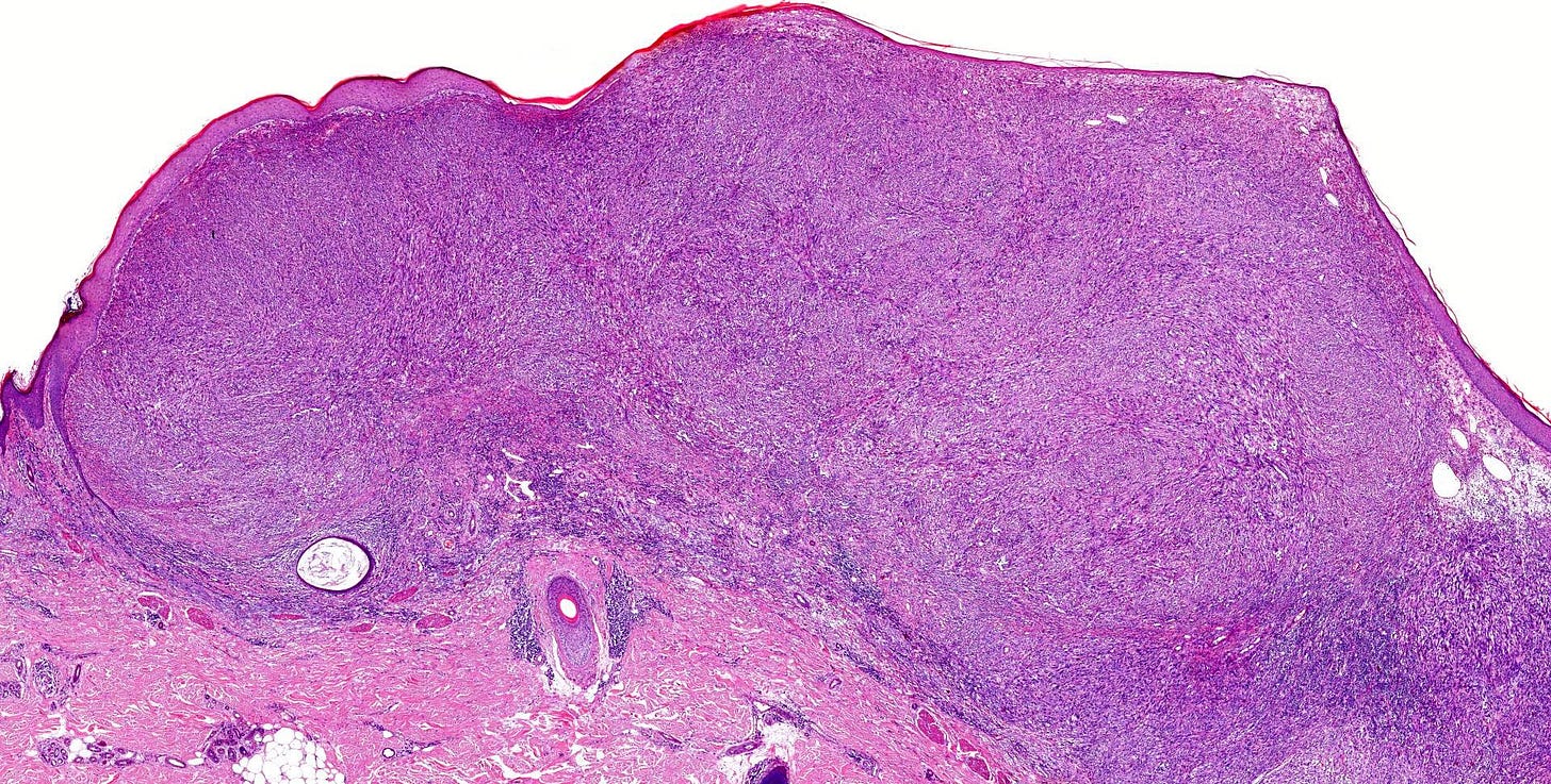
These images are of a variant of melanoma called desmoplastic melanoma, which consists entirely of spindle cells in fibrous tissue without conventional melanoma cells. Contributed by Gregory A. Hosler, M.D., Ph.D.
This is a fine needle aspirate from a mucosal melanoma which shows large rounded cells, some with two nuclei. There are also melanophages, which are inflammatory cells that ingest the melanin pigment. Contributed by Mona Deerwester, M.D.
Index to Nat’s Substack articles
If you like these essays, please share them with others.
Follow me at Substack or LinkedIn or through my Curing Cancer Newsletter .
Follow our Curing Cancer Network on LinkedIn and Twitter. Twice a week we post interesting cancer related images of malignancies with diagnoses.
Latest versions of our cancer related documents:
Strategic plan to substantially reduce cancer deaths
American Code Against Cancer (how you can prevent cancer)
Email me at Nat@PathologyOutlines.com - unfortunately, I cannot provide medical advice.
I also publish Notes at https://substack.com/notes, and would love for you to join me there! Subscribers will automatically see my notes. Feel free to like, reply, or share them around!


