I am now posting interesting cancer images, usually from PathologyOutlines.com, the free online pathology textbook that I own. Click on the links to see the complete topic with its authors and editors.
An MRI of the forearm shows a large lobulated mass infiltrating the muscles.
A gross photo of the tumor (i.e. how it appears after the surgeon removed it), which was cut into 4 sections to better visualize the tumor. The tumor is tan/gray, the fat is yellow, hemorrhage is dark red and most prominent on the left side of the upper right section, and necrosis (i.e. dead tissue) may be at the lower part of the lower right section. When pathologists receive specimens from surgeons, they typically ink them black so they can later determine, under the microscope, if tumor is present at the margin (edges) of the specimen; if so, additional surgery or other treatment may be needed.
Under the microscopic low power, the cancer cells appear in long intersecting fascicles (bundles) - this growth pattern is typical for this tumor.
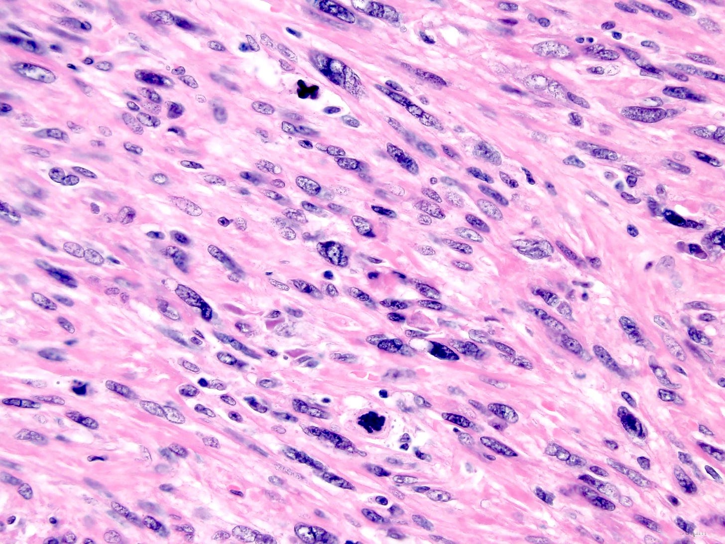
Under the microscopic high power, the tumor cells have pleomorphic nuclei (i.e. they vary from each other), which typically indicates aggressive disease.
Tumor cells show mitotic activity (i.e. cell division) (near the top, left of center; bottom center). This typically indicates malignancy because (a) cells usually divide so slowly that a mitotic figure is rarely present; (b) these mitotic figures are very dark and have an abnormal shape - click here for what a normal mitotic figure looks like.
This is a fine needle aspirate (FNA) of this tumor, which will be interpreted by a cytopathologist, a subspecialty of pathology. The surgeon sticks a hollow needle into the tumor and sucks out cells, which are applied to a slide. This is less invasive for patients than a surgical excision but it may be harder to interpret because the process destroys the three dimensional arrangement of the cells.
Click here for more information on soft tissue leiomyosarcoma.
Index to Nat’s Substack articles
If you like these essays, please share them with others.
Follow me at Substack or LinkedIn or through my Curing Cancer Newsletter .
Follow our Curing Cancer Network on LinkedIn and Twitter. Twice a week we post interesting cancer related images of malignancies with diagnoses.
Latest versions of our cancer related documents:
Strategic plan to substantially reduce cancer deaths
American Code Against Cancer (how you can prevent cancer)
Email me at Nat@PathologyOutlines.com - unfortunately, I cannot provide medical advice.
I also publish Notes at https://substack.com/notes, and would love for you to join me there! Subscribers will automatically see my notes. Feel free to like, reply, or share them around!


Thanks - we will try to do one diagnosis per week.
This is a brilliant short essay.
Very thoughtful of you to draw attention at salient points of leiomyosarcoma of soft tissue.
Images are too good. The nasty cells are jumping out of the screen.