Cell migration in the embryo and fetus (how metastases arise, part 3b-1a)
16 October 2023
Cancer causes death primarily through metastases or the movement of cells from the main tumor mass to other parts of the body.
To understand what causes cancer cells to move from their primary site and how to prevent it, we need to understand how cells normally migrate. This essay discusses embryonic and fetal cells that migrate.
Migration soon after fertilization
Beginning at 5 to 9 days after fertilization, cells in the embryo migrate from the blastula stage to form the gastrula with its three primary germ layers: ectoderm (the outer tube that forms the skin and nervous system), endoderm (the inner tube that forms the GI and respiratory tracts) and mesoderm (the middle layer that forms muscle, bone, the heart and circulatory system, cartilage and other connective tissue).

Stages of Animal Development: Cleavage, Gastrulation, Organogenesis
(general information, nothing about migration)
Cells in these three germ layers then migrate to their target locations in the embryo and fetus and differentiate into distinct cell populations that make up various tissues or organs. However, this is quite complex and what causes the migration is not fully understood.

This embryonic migration of cells is tightly controlled in time and space to maintain homeostasis (i.e. normal physiologic activity); migration defects during the embryonic and fetal period can cause early embryonic death, neurological disorders, congenital heart disease and intellectual disabilities.
Migration of neural cells
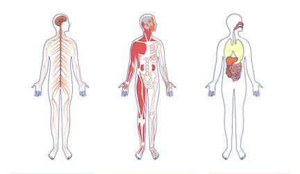
Neural cells migrate to form the nervous system. Cells from the neural crest and their derivatives migrate collectively through the embryo to form:
neurons and glia (supporting) cells,
the medulla (inner portion) of the adrenal gland,
pigmented cells in the skin and
skeletal muscle and connective tissue components of the head.
Migrating neuronal cells

Neuronal migration disorders are described here.
Migration of germ cells to form the testes and ovaries
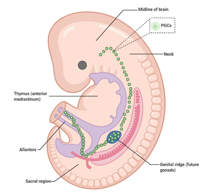
Primordial germ cells migrate as individual cells towards the genital ridges, which become the testes and ovaries

Other cells
Endothelial cells migrate to form blood vessels.
Stem cells migrate to the bone surface and participate in bone formation.
Cessation of migration
After embryogenesis, the migration function is typically shut down but it can be reactivated in adults by cancer risk factors and occasionally by random changes. However, it typically takes years to decades for this reactivation to occur.
Next: Migration in children and adults
Index to Nat’s Substack articles.
If you like these essays, please share them with others.
Follow me on Substack or LinkedIn or through our Curing Cancer Newsletter.
Follow our Curing Cancer Network on LinkedIn and Twitter. Each week we post interesting cancer related images of malignancies with diagnoses.
Latest versions of our cancer related documents:
American Code Against Cancer (how you can prevent cancer)
Email me at Nat@PathologyOutlines.com - Unfortunately, I cannot provide medical advice.
I also publish Notes at https://substack.com/note. Subscribers will automatically see my notes.
Other social media - Tribel: @nat385440b, Threads: npernickmich


