Hank Green, novelist, science communicator and longtime host of YouTube educational videos, recently announced that he was diagnosed with Hodgkin lymphoma and has started chemotherapy. This overview discusses the disease in general, not his specific case.
Lymphomas are a cancer of lymphocytes, a type of white blood cell. There are an extensive variety of lymphomas, generally divided into Hodgkin and non-Hodgkin subtypes. They are also divided into B cell and T/NK cell lymphomas.
Hodgkin lymphoma is a B cell lymphoma. Most cases are classified as classic Hodgkin lymphoma, which has 4 subtypes: nodular sclerosis (the most common subtype), lymphocyte rich, mixed cellularity and lymphocyte depleted. The nonclassic type is called nodular lymphocyte predominant B cell lymphoma or nodular lymphocyte predominant Hodgkin lymphoma.
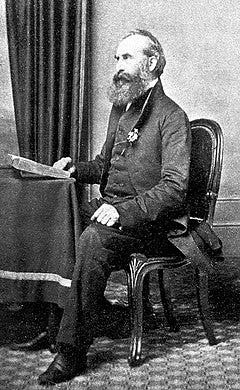
Dr. Thomas Hodgkin, source: Wikipedia
Hodgkin lymphoma (HL) used to be called Hodgkin’s disease (named for Dr. Thomas Hodgkin) because physicians did not understand its origin, partly because it causes tumors composed of very few malignant cells surrounded by large numbers of nonmalignant inflammatory cells.
Epidemiology (who gets it):
The epidemiology varies by the subtype; for the nodular sclerosis subtype (70% of classic HL), patients are usually ages 15 - 35 years, living in urban areas and having high socioeconomic status. It is more common in females than males.
Factors associated with increased risk include a history of infectious mononucleosis or HIV infection or a family history of classic HL, particularly in siblings. However, most patients have no risk factors.
Clinical features
Most patients present with painless lymphadenopathy (enlarged lymph nodes). 30 - 40% of patients present with B symptoms (fever, night sweats, weight loss) or pruritis (itching).
Before treatment, patients are staged, which means they undergo studies to determine the extent of disease. Treatment varies by stage. Fortunately, Hodgkin lymphoma has a high cure rate (80 - 90%).
Pathophysiology: How does Hodgkin lymphoma arise?
The malignant cells in Hodgkin lymphoma are called Hodgkin and Reed-Sternberg cells (Hodgkin cells if they have one nucleus and Reed-Sternberg cells if >1). They originate as B lymphocytes (B cells), which may be activated in the germinal center region of the lymph nodes, spleen or related tissue through a multistep process to become plasma cells, which produce antibodies and have limited lifespans. However, this process may be disrupted in Hodgkin lymphoma (at the preapoptotic germinal center step) to produce Hodgkin or Reed-Sternberg cells. These cells have malignant properties and, unlike normal lymphocytes or plasma cells, they live indefinitely (Blood 2000;95:1443).
Reed-Sternberg cells secrete cytokines (substances that have effects on other cells), which recruit nonmalignant inflammatory cells to the area and create the bulk of the tumor mass. These inflammatory cells, although not malignant themselves, also produce cytokines, which support the survival and proliferation of the Reed-Sternberg cells.
I have suggested (but not proven) that Hodgkin lymphoma may be due to a “runaway” immune system. The immune system typically maintains a delicate balance between activating and dampening forces which may be disturbed by antigens (substances, typically foreign to the body, that trigger an immune response), random or other trivial events. Over time, these disturbances may propagate throughout the immune system, reinforce the process and ultimately create premalignant and malignant cells.
What do the enlarged lymph nodes look like?
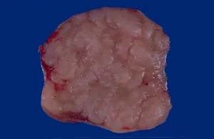
The lymph nodes of Hodgkin lymphoma typically are enlarged and nodular with a thickened capsule (outer covering). The nodular sclerosis subtype has prominent fibrotic bands:


Enlarged lymphoma nodes with Hodgkin lymphoma: Sources: left, middle, right.
In comparison, a normal lymph node is smaller and homogeneous, with no nodularity:
Normal lymph node for comparison, contributed by Patricia Tsang, M.D., M.B.A.
What is the microscopic (histologic) appearance?
There is total or partial effacement (destruction) of the normal lymph node architecture.
The nodular sclerosis subtype shows nodularity, dense collagen bands and a thickened capsule.
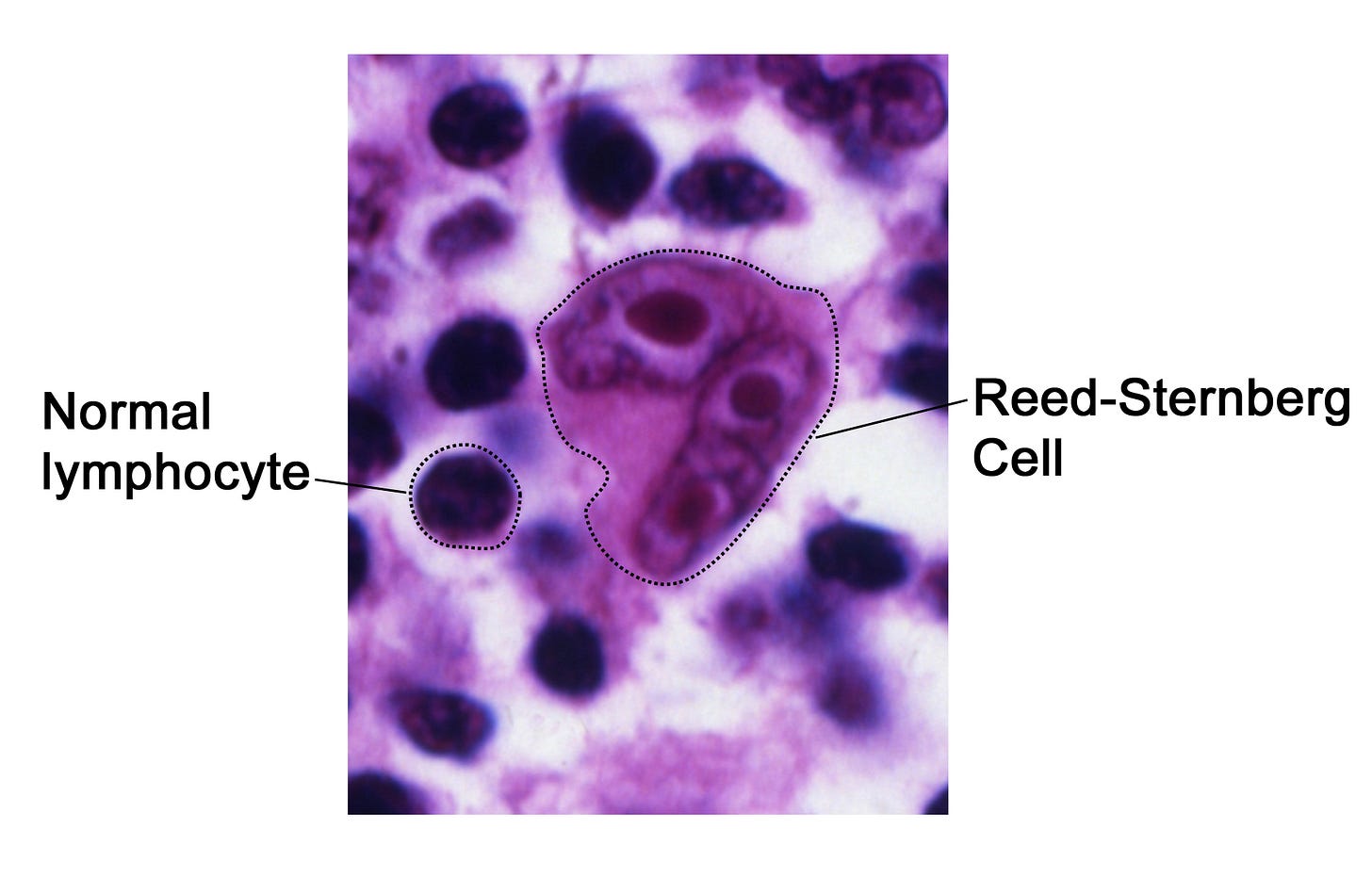
The Reed-Sternberg cell is large (up to 100 microns) with a bilobated to multilobated nucleus, a prominent eosinophilic (pink/red) nucleolus and ample amphophilic (between blue and pink/red) cytoplasm. They may be rare.
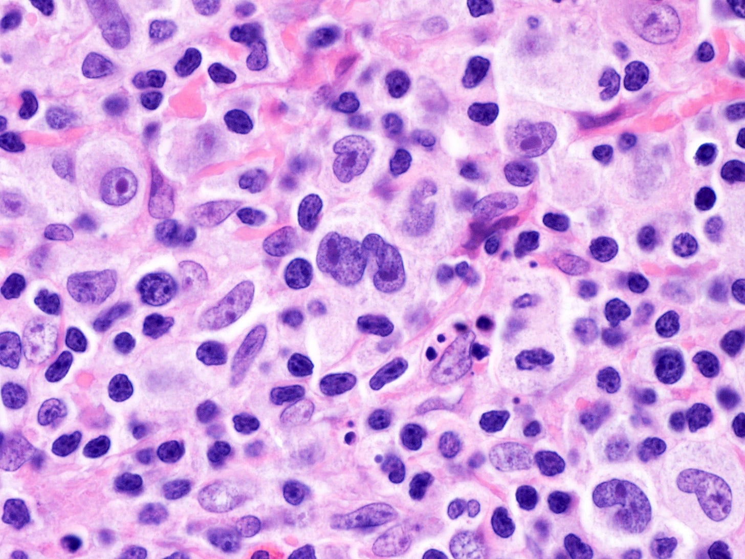
The background has inflammatory cells, which varies based on the subtype (Br J Haematol 2019;184:45):
Source: Wikipedia
Reed-Sternberg cells have 2 lobes and often have multiple nuclei, Source #1, #2
Of note, biopsies in infectious mononucleosis show cells that look very similar to Reed-Sternberg cells, which is why getting a clinical history is often so important to pathologists.
Index to Nat’s Substack articles
If you like these essays, please share them with others.
Follow me on Substack or LinkedIn or through our Curing Cancer Newsletter .
Follow our Curing Cancer Network on LinkedIn and Twitter. Twice a week we post interesting cancer related images of malignancies with diagnoses.
Latest versions of our cancer related documents:
American Code Against Cancer (how you can prevent cancer)
Email me at Nat@PathologyOutlines.com - unfortunately, I cannot provide medical advice.
I also publish Notes at https://substack.com/note. Subscribers will automatically see my notes.


