Few men have mammograms - I had 4 as of September 2023. Here is my story:
In early 1984, I started treatment for Hodgkin lymphoma, a previously lethal form of cancer that had spread throughout my abdomen and pelvis (see my prior post). I received radiation therapy, the standard treatment at the time, which caused all the cancer to disappear. I am now considered cured.
In Hodgkin lymphoma patients, there is an increased risk of breast cancer in women or girls who, like me, received mantle field radiation therapy (which includes the chest), but there is little data for men and boys.
Two years ago, I had right sided rib pain and saw an ER doc who ordered Xrays, which were normal. He prescribed Motrin, which worked immediately (for the diagnosis of costochondritis). Subsequently, I mentioned this to my internist, who suggested I see a breast physician, “out of an abundance of caution”. The oncologist ordered a mammogram.
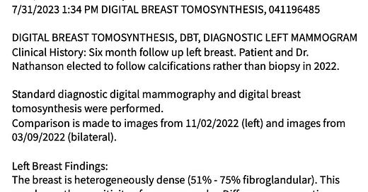
The right breast was completely normal but the left breast had “abnormal calcifications” and the radiologists recommended a biopsy. But, as a non-overweight man, there was so little breast tissue to biopsy that a simple mastectomy (i.e. removing all breast tissue) would have been required to get sufficient tissue. In addition, the oncologist did not think the calcifications looked that suspicious. In my experience as a pathologist, radiologists sometimes “overcall” lesions. In any event, we decided on follow-ups instead of surgery, and to date, the calcifications are “stable” (i.e. they have not changed) over subsequent mammograms. This is the most recent report:
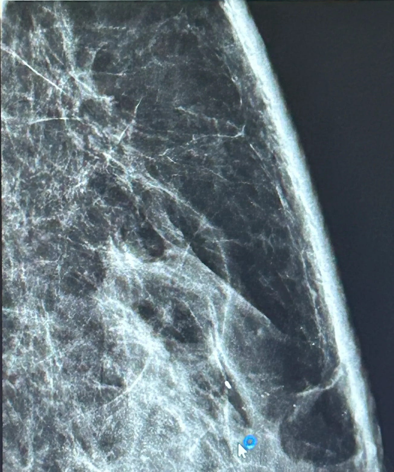
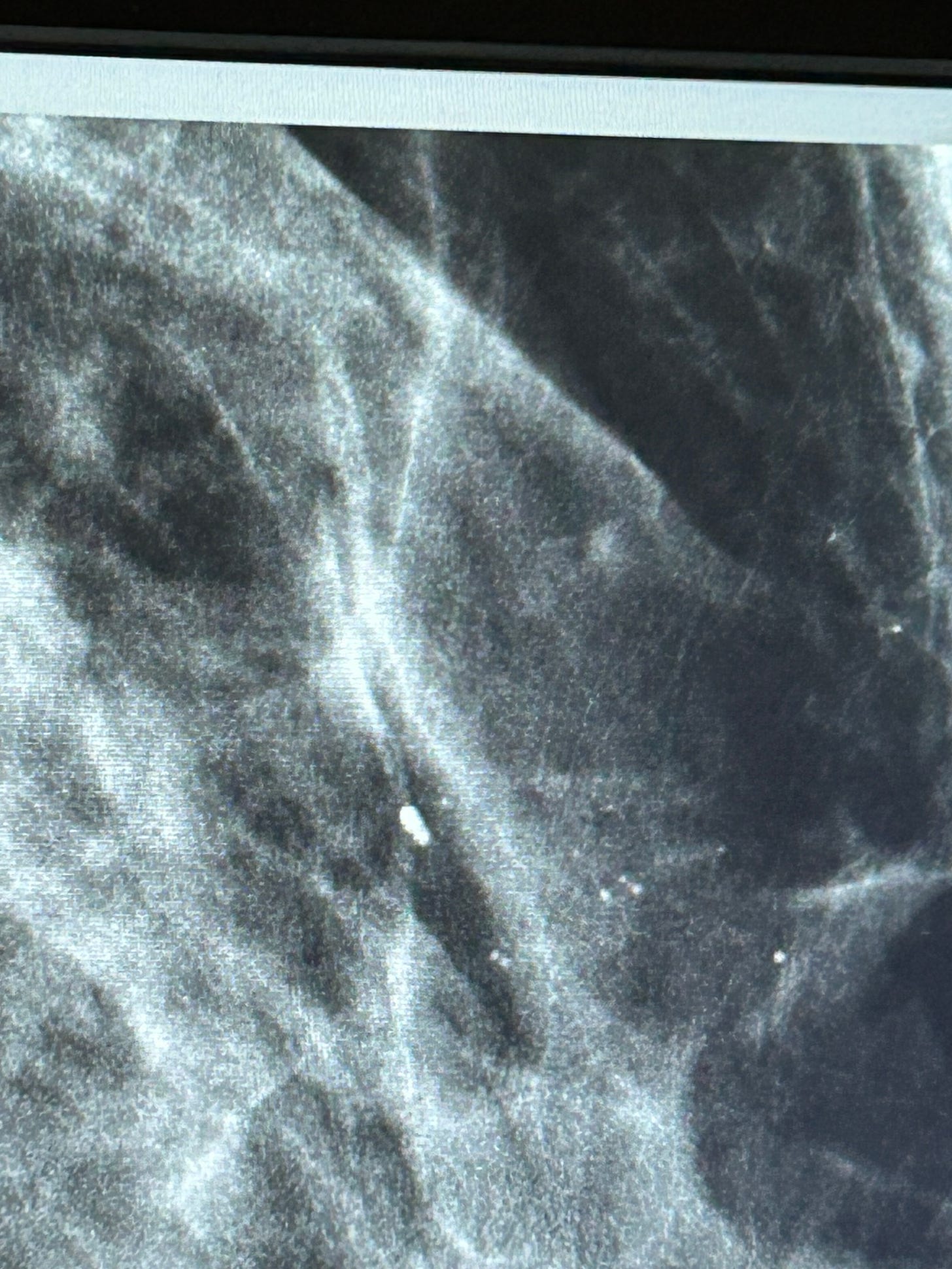
The mammographic images are below. They were taken from the Xray machine with my iPhone after a discussion with the radiologist. I also reviewed them with a radiologist friend. Although the radiology department gave me images on a CD, I was unable to read them (it may have required special software). Below, the small white circular “dots” near the arrow towards the bottom are the calcifications; the white linear streaks are fibrous tissue and can be ignored.
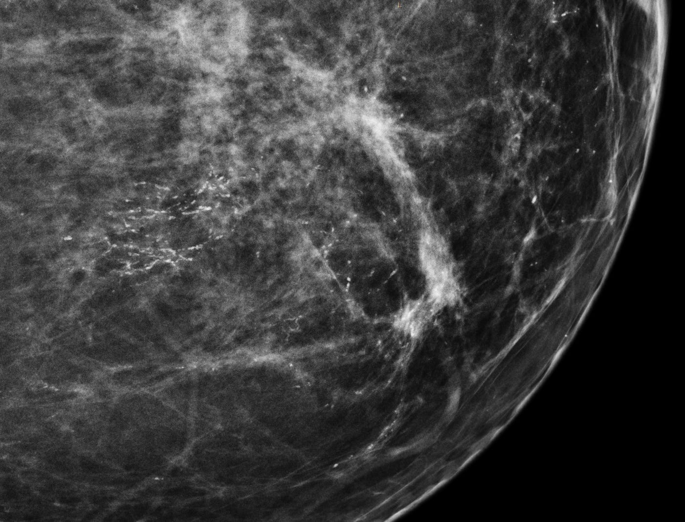
For comparison, here are microcalcifications for breast ductal carcinoma in situ (DCIS), a preinvasive malignant lesion that does require treatment.

In DCIS, the microcalcifications are much denser.
I will have another mammogram in 6 months.
Index to Nat’s Substack articles\
If you like these essays, please share them with others.
Follow me on Substack or LinkedIn or through our Curing Cancer Newsletter.
Follow our Curing Cancer Network on LinkedIn and Twitter. Each week we post interesting cancer related images of malignancies with diagnoses.
Latest versions of our cancer related documents:
American Code Against Cancer (how you can prevent cancer)
Email me at Nat@PathologyOutlines.com - Unfortunately, I cannot provide medical advice.
I also publish Notes at https://substack.com/note. Subscribers will automatically see my notes.
Other social media - Tribel: @nat385440b, Instagram / Threads: npernickmich


